Tracking intrinsically fluorescent drugs and autofluorescent biomarkers
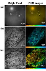
When a drug is intrinsically fluorescent, like doxycycline, minocycline, etc, things are somewhat easier. You can use tunable lasers or a spectrometer to characterise the absorption spectra of these drugs, and by using a prism-based detection approach we remove the need to purchase unique emissions filters and can instead tune our emission gates to whatever wavelengths we choose. If you have a pulsed laser and can do time-correlated single photon counting (TCSPC) for fluorescence lifetime imaging microscopy (FLIM), that allows you to measure the decay curve of each fluorophore in the emission window, thereby enabling mapping of the drug independent from tissue autofluorescence background (Figure 1).

Figure 1: Bright field and FLIM images of (a) dried minocycline, (b) an untreated sebaceous gland, and (c) a sebaceous gland treated with minocycline. [https://www.ncbi.nlm.nih.gov/pmc/articles/PMC6033575/].
By using a tunable multiphoton laser we can probe even deeper into tissue using near infrared excitation and eliminating the need for tedious tissue sectioning. Furthermore, with that same multiphoton microscope we can use second harmonic generation for label-free imaging of large non-centrosymmetric structures such as collagen, microtubules and muscle myosin, which may serve as biomarkers for a drug’s efficacy.
Characterising nanoparticle-based assays and nanoparticle drug delivery vehicles
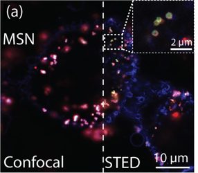
Many pharmaceutical researchers may also have the need to characterise nanosized drug delivery vehicles that are smaller than the diffraction limit of traditional confocal microscopes. For this, we can utilise super-resolution microscopy techniques like STED and STORM, which enable fluorescence imaging at the scale of ~20-40nm (nanoscopy). In our facility, STED can be done in combination with FLIM, called tau-STED, meaning we can also get lifetime information at the nanoscale. While very useful for characterising the structure and distribution of nanomaterials in cells, tissues, or in microfluidic chip platforms, the downside of imaging at the nanoscale is that it can take a long time to image over large areas (Figure 2).
Figure 2: Interactions of protein corona-coated nanoparticles with cells. Confocal and STED images of mesoporous silica nanoparticles (MSNs) after incubation with cells followed by fixation. Zoom-in STED images show nanoparticles internalised through cell membrane
Visualising drug effects in whole organs and small animals
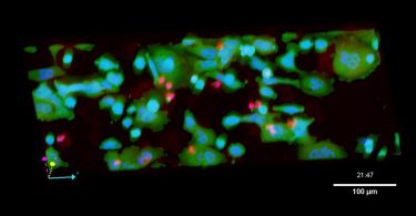
For macroscale characterisation and monitoring system-wide drug effects of porous nanoparticles, lysosomes, PLGA-encapsulated drugs and more, we turn to techniques like spinning disk confocal, lightsheet and selective plane illumination microscopy (SPIM), which enable researchers to see the pharmacokinetics of a drug in real time and in situ. Whether that be in a tissue construct, organoid, or whole living animal, stage-top incubation systems allow us to dynamically image large volumes of tissues and small animal models over the course of days with millisecond temporal resolution (Figure 3).
What’s next on the horizon?
In the future we hope to build correlative systems, which would allow us to mix and match several of these techniques with each other; for example, FLIM with light sheet. We also hope to develop new correlative techniques such as combining super-resolution nanoscopy with electron microscopies or combining fluorescence with molecularly sensitive spectroscopic techniques such as Raman scattering and MALDI imaging.
Author Bios:
Dr Haley Marks
Haley is a biomedical engineer with interests in nano-biosensor research, translational medicine and optics education, and is a past recipient of the Whitaker International and SPIE Franz Hillenkamp fellowships. As a project scientist at CNSI in UCLA, Haley serves as a technical expert in the Advanced Light Microscopy and Spectroscopy centre, providing light microscopy training and imaging services to ALMS users of all backgrounds. She also develops customised experimental strategies and imaging workflows, optimises ALMS’s existing super-resolution and high-speed optical methods, and disseminates computational imaging techniques.
Dr Laurent A. Bentolila
Laurent is a Senior Research Scientist, Lecturer and Director of the Advanced Light Microscopy/Spectroscopy Laboratory at the California NanoSystems Institute at UCLA. Laurent’s long-standing research interest focuses on the application of nanotechnology and advanced light microscopy techniques to biology and medicine. He is the recipient of several awards including the European Molecular Biology Organization and the Burroughs Welcome Fund.
Brian Jeong
Brian is a research & development engineer with a background in biophysics. His interdisciplinary skillset, combined with experience in hardware configurations and software implementations, enables him to devise and optimise customised imaging solutions for diverse research project goals. His current research focus is the development of microscopy modalities to capture volumetric dynamics, as well as data analytics for fluorescent microscopy and AI personalised medicine.